
Anatomy of your ear
Your ear is responsible for both hearing and balance. Knowing a bit more about the structure and function of the different parts of your ear can help you understand your diagnosis and treatment. Your ear is made up of three parts: outer, middle, and inner.
Outer ear
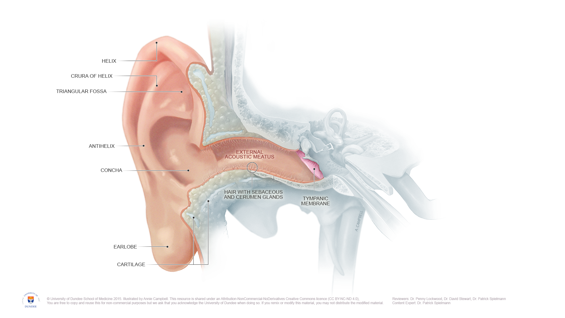
The outer ear is the visible part of your ear which is called the auricle or pinna, and the ear canal. This part of your ear allows sound waves to be transmitted to the eardrum.
Problems that can occur in the outer ear include otitis externa (swimmer’s ear or outer ear infection) and cerumen impaction (blockage of ear wax).
Middle ear
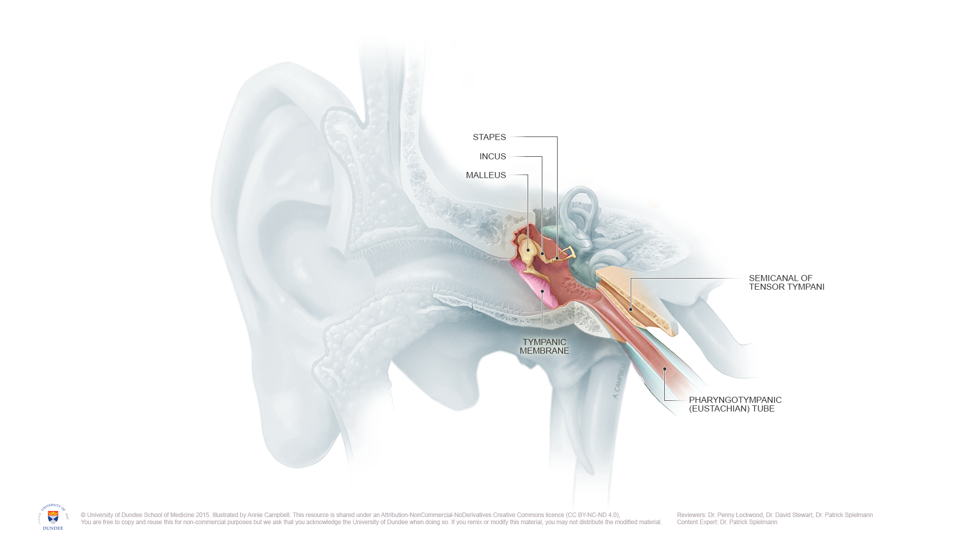
The middle ear includes your eardrum, also known as the tympanic membrane, and the air-filled space behind the eardrum.
This is the location of your bones of hearing - called the ossicles. The malleus (“hammer”), incus (“anvil”), stapes (“stirrup”) are tiny bones that transmit the vibrations of the ear drum to the inner ear.
The middle ear space is connected to the eustachian tube. The eustachian tube equalizes pressure with your environment by allowing air to enter the middle ear. It is connected to the nasopharynx - the back of your nose and top of your throat.
Problems related to the middle ear include otitis media (middle ear infection), tympanic membrane perforation (hole in the eardrum), cholesteatoma, otosclerosis, ossicular chain disruption (change in alignment of the ossicles), and eustachian tube dysfunction.
Inner ear
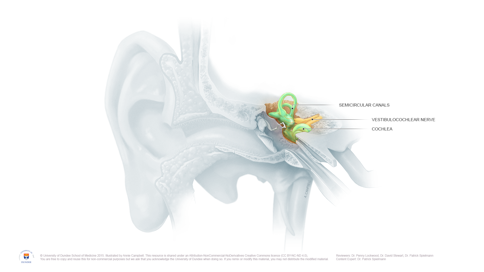
Your organs of hearing and balance are in your inner ear. The inner ear is located within the temporal bone of your skull.
The cochlea is responsible for hearing. Sound waves cause the eardrum to vibrate, and these vibrations are passed to the ossicles. The ossicles send amplified vibrations to the cochlea. In the cochlea, the vibrations are converted into electrical signals, which are sent to the brain by the auditory/cochlear branch of the vestibulocochlear nerve. Your brain then interprets the nerve signals as sound.
The vestibular organs are responsible for balance. This part of your ear is also known as the labyrinth. There are two parts to your vestibular organs, the otoliths and the semicircular canals. The vestibular organs sense head motion, and send electrical signals about balance and movement to the brain via the vestibular branch of the vestibulocochelar nerve.
The otoliths are located in the vestibule, and sense linear motion and head tilt. There are two parts to your otoliths, the utricle and the saccule. Your utricle senses horizontal motion. Your saccule senses vertical motion.
The semicircular canals sense rotational motion. You have three semicircular canals that are oriented at 90 degree angles to each other. They are named for their location: anterior (also known as superior - at the front or top), posterior (at the back), and lateral (to the side).
The vestibulocochlear nerve (cranial nerve eight) sends signals about hearing and balance from your inner ear to your brain.
Problems related to the inner ear include vestibular neuritis and labyrinthitis, benign paroxysmal positional vertigo (BPPV), Meniere’s Disease, superior semicircular canal dehiscence, perilymphatic fistula, medication-related ototoxicity, and vestibular schwannoma.
Inner ear disorders often result in symptoms of vertigo, dizziness, imbalance, and hearing loss. Vestibular rehabilitation is effective at evaluating and treating inner ear balance problems.
Our physiotherapists have expert knowledge of inner ear anatomy and function. Want to know if vestibular rehab could help you? Get in touch to talk to our team!